
Patellar instability surgery
Surgery for patellar instability encompasses all procedures designed to treat episodes of patellar dislocation or subluxation. It is recommended when instability persists despite conservative treatment or in cases of recurrence.
The objective is to permanently stabilize the kneecap, reduce pain, and limit the risk of further dislocations. The surgical technique is adapted to the anatomical abnormalities identified during the pre-operative assessment.
Patellar instability surgery
Surgery for patellar instability encompasses all procedures designed to treat episodes of patellar dislocation or subluxation. It is recommended when instability persists despite conservative treatment or in cases of recurrence.
The objective is to permanently stabilize the kneecap, reduce pain, and limit the risk of further dislocations. The surgical technique is adapted to the anatomical abnormalities identified during the pre-operative assessment.
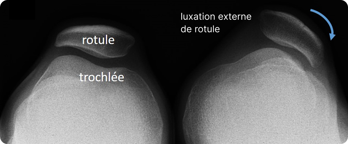
When is surgery for patellar instability necessary?
Several situations may lead to considering surgical management.
Potential rotational instability
Certain anatomical abnormalities can promote poor centering of the kneecap during knee movements and lead to repeated episodes of subluxation and/or a feeling of instability.
Recurrent patellar dislocations
After a first episode of dislocation, some patients suffer from recurrences that cause functional discomfort and instability in daily life or during sports activities.
Chronic knee pain
Patellar pain, locking episodes, or isolated sensations of instability may lead to the proposal of compensatory surgery.
After failure of medical treatment
A treatment combining rehabilitation, muscle strengthening, and activity modification is generally offered as a first-line approach. When this does not provide sufficient improvement, surgical intervention may be considered.
How is the diagnosis established?
The diagnosis is based on a specialized consultation combining a clinical examination and imaging workup.
X-rays make it possible to study the position of the patella and the alignment of the lower limb. Depending on the case, additional examinations may be prescribed:
- scanner specifically making it possible to measure factors favoring instability: TT-TG, patellar tilt, patellar height, and trochlear and patellar dysplasia.
- MRI of the knee ;
- specific radiographs of the patellofemoral joint.
These tests make it possible to identify the factors responsible for the instability and to define the most appropriate surgical strategy.
How does the pre-operative care take place?
Before the procedure, an assessment is carried out in order to plan the surgical treatment. It generally includes an anesthesia consultation as well as the imaging exams necessary for the evaluation of the bony, ligamentous, and cartilaginous structures of the knee, or any other consultation made necessary.
What does the procedure involve?
The procedure is performed under general or loco-regional anesthesia (spinal anesthesia). The surgical technique depends on the abnormalities found during the pre-operative assessment and may combine several procedures to improve patellar stability.
Tibial tubercle osteotomy
It may consist notably in performing an osteotomy of the anterior tibial tuberosity, onto which the patellar tendon inserts, making it possible to modify its position on the tibia and thus improve the centering of the extensor mechanism and particularly of the patella.
External patellar retinaculum release
Another frequently performed procedure is the lateral retinacular release, which consists of releasing the structures located on the outer side of the patella when they exert excessive traction pulling the patella toward the lateral edge of the knee.
These procedures can be combined with one another or supplemented by other techniques depending on the patient's needs: strengthening of the vastus medialis or the medial patellofemoral ligament (MPFL).
Mandatory instructions before surgery
To guarantee the success of the procedure and minimize the risk of infection as much as possible, certain strict instructions must be followed before you are admitted to the hospital:
Smoking cessation
Since smoking massively increases the risks of infection and delayed healing, patients are strictly required to stop smoking at least one month before the procedure and for the three months that follow.
Skin preparation
The day before and the morning of the procedure, you must take a shower using an antiseptic or disinfectant soap. Please note that shaving the knee is not recommended; if hair removal is necessary, it must be done with hair clippers or a depilatory cream to avoid any micro-cuts.
Reporting allergies
You must absolutely inform your surgeon and the anesthesiologist if you have allergies to antibiotics, as well as to metals (such as nickel, chromium, or cobalt) if you have ever reacted to contact with jewelry, a watch, or a belt buckle.
How does post-operative follow-up work?
Rehabilitation begins quickly after surgery in order to progressively recover knee mobility and restore joint function. A brace is often necessary to wear for 30 to 45 days; driving a car can only be done after its removal.
Resumption of weight-bearing is authorized according to the surgeon's instructions (most often immediate) and depends on the procedures performed. Supervised rehabilitation is essential to optimize the functional outcome.
The resumption of sports activities is gradual and takes place after medical clearance, respecting the consolidation and recovery periods, generally at least 3 months.
The pre-operative notes
Any surgery carries the risk of difficulties or complications which, even if uncommon, must be known and taken into account in the surgical decision. Please click below to read the information corresponding to the procedures described above.
Tibial tubercle osteotomy
External patellar retinaculum section